
Clinical Physiology of Circulation
ISSN 1814-6910 (Print)
ISSN 2410-9134 (Online)


Chief Editor
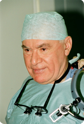
Leo A. Bockeria, MD, PhD, DSc, Professor, Academician of Russian Academy of Sciences, President of Bakoulev National Medical Research Center for Cardiovascular Surgery
Sort by
Diagnostics of progression predictors of abdominal aortic aneurysm by computed tomographic angiography

Authors:
Company:
Bakoulev National Medical Research Center for Cardiovascular Surgery, Moscow, Russian Federation
E-mail: Сведения доступны для зарегистрированных пользователей.
DOI:
UDC: 616.136-007.64-073.75
Link: Clinical Physiology of Blood Circulaiton. 2023; 2 (20): 140-154
Quote as: Getsadze G.G., Aslanidis I.P., Dorofeev A.V., Arakelyan V.S., Mamalyga M.L., Fedorchenko P.V. Diagnostics of progression predictors of abdominal aortic aneurysm by computed tomographic angiography. Clinical Physiology of Circulation. 2023; 20 (2): 140–54 (in Russ.). DOI: 10.24022/1814-6910-2023-20-2-140-154
Received / Accepted: 30.03.2023 / 21.04.2023
DownloadAbstract
Objective – identification of predictors of abdominal aortic rupture and their threshold levels in abdominal aortic aneurysm (AАA) progression on the basis of MSCT study.
Material and methods. The study included 70 patients with infrarenal AAA divided into 3 groups: group 1 – with stable wall, group 2 – with unstable wall, group 3 – with AAA rupture. For each group, the following parameters were estimated at different levels: 1) maximum diameters and areas of: vessel, lumen, and wall at the level of the proximal AAA neck, at the level of maximum AAA dilation, and at the level of the terminal aortic section; 2) aneurysm extent; 3) ratio of aneurysm diameter to normal aortic diameter; 4) ratio of thrombosis area to aneurysm area; 5) tangential tension of the aortic wall; 6) degrees of calcification of the aneurysmal segment and the entire abdominal aorta. CT images were analyzed retrospectively on an image storage and transmission system workstation (Philips). Statistical analysis was performed using IBM SPSS program (version 26).
Results. According to the study wall instability occurred when AAA area increased 35% (P = 0.017) and tangential stress increased 1.3 times (P = 0.007). The ratio of thrombosed wall area to aneurysm area was 72%. There was no change in lumen area. ROC analysis showed thresholds of unstable wall: maximum aneurysm size of 58.7 mm; aneurysm area of 24.5 cm2, wall area of 15.4 cm2; ratio of thrombosed wall area to aneurysm area of 0.66. Further progression of AАA compared with stable aneurysm leads to a 1.55–fold increase in its extent at rupture (P = 0.004), a 58% increase in proximal neck vessel area (P = 0.019), and a 19% increase in lumen area (P = 0.044). Vessel area 5.7 cm2, lumen 4.1 cm2, and maximum diameter 28.3 mm can be considered threshold values according to ROC analysis at the level of the proximal neck. AAA rupture was defined as 2.9-fold (P < 0.001) increases in AAA area, 4.8-fold (P < 0.001) increases in wall area, and 1.7-fold increase in lumen area (P = 0.029). The threshold values for maximum AAA size were 7.6 cm, area was 34.4 cm2, and wall area was 17.4 cm2. Dilation of the terminal aorta can also be considered as a predictor of AAA rupture. Patients’ examination showed a 2.75-fold (P = 0.019) increases in terminal area, 4.5-fold (P = 0.010) increases in wall area, and 2.1-fold (P = 0.049) increases in lumen area. At aortic aneurysm rupture according to ROC analysis threshold vessel area in the terminal section was 14.7 cm2, wall area – 7 cm2, thrombosis thickness – 11.9 mm.
Conclusions. Analysis of the study showed that the threshold predictors of AAA rupture are maximum AAA diameter 7.6 cm, AAA area 34.4 cm2, AAA wall area 17.4 cm2, proximal neck area 5.7 cm2, vessel area, wall area and thrombosis thickness in the terminal aorta 14.7 cm2, 7 cm2, 11.9 mm respectively.
About Authors
- Gela G. Getsadze, Researcher, Radiologist of Department of Cardiovascular Radiology Diagnostics; ORCID
- АIrakliy P. Aslanidis, Dr. Med. Sci., Professor, Deputy Director, Head of Department of Nuclear Diagnostics; ORCID
- Aleksey V. Dorofeev, Cand. Med. Sci., Head of X-Ray Diagnostic Department, CT and MRI; ORCID
- Valeriy S. Arakelyan, Dr. Med. Sci., Professor, Head of Department of Arterial Pathology Surgery; ORCID
- Maksim L. Mamalyga, Dr. Med. Sci., Senior Researcher of Department of Surgical Treatment of Ischemic Heart Disease; ORCID
- Polina V. Fedorchenko, Researcher, Radiologist of Department of Cardiovascular Radiology Diagnostics; ORCID
If you found mistakes, select text and press Alt+A