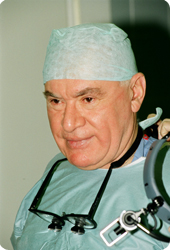
Leo Antonovich BOCKERIA,
Academician of RAS and RAMS,
President of Bakoulev National Medical Research Center for Cardiovascular Surgery
Chief Editor of the Center’s Publishing House
Cardiac surgeon; scientist and researcher; pedagogue and science organizer; doctor of medicine (1973); professor (1982); academician of RAMS (1994), academician of RAS (2011); Honored Scientist of the Russian Federation (1994); Lenin Prize (1976), State Prize of the USSR (1986), State Prize of the RF (2002) and the Government of the RF Prize (2003) laureate.
In 1994 Lео A. Bockeria was elected the Director of A.N. Bakoulev Scientific Center for Cardiovascular Surgery (SCCVS). In 1998 he became at the same time the Director of the Center for Surgical and Interventional Arrhythmology under the Ministry of Healthcare of the Russian Federation (MHС RF).
Since 1994 he has been the Head of the Department of Cardiovascular Surgery at the Russian Medical Academy of Postgraduate Education (MHC RF). Since 1995 he has been the Head of the Department of Cardiovascular Surgery № 2 (which was founded by him personally) at Moscow Medical Academy (now the First Moscow Medical University) named after I.M. Sechenov (MHC RF).
Since 2003 he has led the Department of Cardiovascular Surgery and Interventional Cardiology at Moscow State Medical and Stomatological University named after A.I. Evdokimov (MHC RF).
By his initiative the Training and Research Center ‘Modern Medical Technologies’ successfully functions, running annually 4–6 courses of postgraduate qualification improvement for senior specialists.
Lео A. Bockeria perfoms the entire known plurality of heart operations in the most various pathologies: 3–6 operations daily, that is 700–900 operations with extracorporeal circulation a year.
Since 1996 he has been the Chief Cardiac Surgeon of the MHС RF.
He is the author and co-author of more than 3700 scientific publications including more than 250 books, more than 100 inventions and utility models, more than 300 computer programs and databases, some of which are registered abroad, designed for various problems of cardiоvascular surgery, cardiology, medical education and medical science organizing.
Lео A. Bockeria is the author of the unique works dedicated to the theoretical foundation and clinical use of the method of hyperbaric oxygen therapy in heart and vessels surgery. He is the leading expert in diagnostics and surgical treatment of the heart rhythm and conduction disorders (especially tachyarrhythmias) including various combinations of heart arrhythmias with congenital and acquired defects as well as malformations, coronary disease.
He also initiated the development of another new section of cardiac surgery in Russia – minimally invasive heart surgery. He made a great contribution to the solution of the problem of coronary heart disease surgical treatment.
His role in the development of new approaches to the surgical treatment of terminal heart failure is great. The development of operations in a heavy category of cardiac patients with different forms of cardiomyopathies is his achievement. He was the first to amplify the concept of dynamic cardiomyoplasty, including that in children. Lео A. Bockeria performed the first implantation of artificial ventricle in Russia and after a long break initiated the resumption of heart transplantations in the Center.
It is by his initiative that the advanced technologies of diagnostics and reconstructive surgery of the ascending aorta and aortic arch disorders have been introduced into clinical practice.
Besides, Lео A. Bockeria ensured the development of another trend in modern medical science – creative cardiology, which determinates the necessity of the constructive cooperation of physicians representing different specialities: cardiologists, cardial surgeons, functional diagnosticians, experts in fundamental and applied disciplines.
Under the leadership of Lео A. Bockeria the new highly technological principles of prevention and treatment of patients with cardiovascular pathologies are worked out and introduced into clinical practice in the Center – namely using the methods of gene and cell therapy.
Academician Lео A. Bockeria conducts priority researches to create bioprosthetic heart valves for valve defects correction. A study of the effectiveness of myocardial protection in infants under the age of one year using new intracellular cardioplegic solution ‘Bockeria – Boldyreva’, created in the SCCVS under Bockeria’s guidance, is continued.
Lео A. Bockeria is actively engaged in methodology of medical science and pedagogical activities. He created the largest in Russia cardiac school and trained more than one generation of physicians – heart surgeons, cardiologists, resuscitators and specialists of other related professions.
Lео A. Bockeria is a scientific supervisor of 350 candidate dissertations and an advisor of more than 100 doctoral thesises. He is the creator of A.N. Bakoulev SCCVS Publishing House with its own printing office, the founder and the Chief Editor of such journals as ‘Annals of Surgery’, .‘The Bulletin of A.N. Bakoulev SCCVS Cardiovascular Diseases’, ‘Clinical Physiology of Blood Circulation’, ‘Annals of Arrhythmology’, ‘Creative Cardiology’, information collection ‘Cardiovascular Surgery’; the Chief Editor of ‘Children’s Heart and Vascular Diseases’ journal.
For his vigorous activities and contribution to the Russian healthcare system Lео A. Bockeria was aworded titles and prizes of the highest level. He is a laureat of Lenin Prize (1976), two State Prizes (1986 – USSR, 2002 – RF), the Russian Federation Government Prize (2003). For prominent achievements he was aworded The Order ‘For Merit to the Fatherland’, Grade III (1999), II (2004) and IV (2010); Georgian Order of Honor and Merit (1999); The Order of Sergius of Radonezh, Grade II (2001). The Russian Biographical Institute repeatedly gave L.A. Bockeria the honorary title ‘Person of the Year’, and in 2000 – ‘Person of the Decade’ in the category of medicine. In 2002 he got the title of the ‘Man of Legend’; the nationwide prize ‘Russian National Olympus’, established by the Government, the Union of Industrialists and the ‘Third Millennium’ fund. In 2003 as one of the world's leading cardiac surgeons Lео A. Bockeria got the international ‘Golden Hippocrates’ aword. In 2004 he was aworded the RAS Premium ‘Triumph’ in the nomination ‘Medicine – the Science of Life’ and the order ‘Maecenas’ of the charity fund ‘Maecenases of the Century’ for the outstanding сontribution to the revival and prosperity of the world, the greatness of the soul, the selfless generosity; in 2004 and 2005 – golden honorary badge ‘Public Recognition’ of the homonymous National Fund, National Civil Committee for Cooperation with the Law Enforcement, Legislative and Judicial Bodies, independent organization ‘Сivil Society’ for the great personal contribution to the developement of national medicine, for performing of the unique cardiosurgical operations using the latest medical technologies, which saved hundreds of children’s and infants’ lives, for long and fruitful scientific-practical, pedagogical and educational activities, for the active citizenship.
In 2006 Lео A. Bockeria was awarded Order of Honor ‘Public Recognition’; diploma of the Presidium of the Parliament of the Peoples of Russia ‘for the salvation of many lives, outstanding knowledge, leadership qualities, for unique personal qualities – nobleness, courage, sense of duty, the ability to cherish the honor and dignity, to keep the word and do the business, as well as for the faith in great future of the Fatherland’; Golden Medal for outstanding сontribution to the education in Russia; Golden Star ‘Honor, Pride and Glory of Russia’.
In 2008 Lео A. Bockeria got the medal ‘for practical contribution to strengthening of the nation's health’; Order of Honor with the awarding of the title ‘Support of Honest Business’ and the honorary title ‘A Distinguished Surgeon of Modern Times’. In 2009 his great contribution to the science and Russian healthcare was marked by awarding him the A.N. Kosygin Prize ‘For Great Achievements in the Development of Russian Economy’ and Moscow city Award in medicine for the development and introduction into clinical practice of the new biological heart valve ‘Bioglis’.
Lео A. Bockeria is a member of the American Association for Thoracic Surgery (1991), Board member (1992) and member of the Presidium (since 2003, consul) of the European Association for Cardio-Thoracic Surgery, Board member of the European Association for Cardiovascular Surgery, Scientific Board member of Monaco Cardio-Thoracic Center (1992), the member of the Serbian Academy of Sciences and Arts (1997), Honorary Member of the American College of Surgeons (1998), the Academician of the Ukraine AMS, Honorary Professor of M.V. Lomonosov Moscow State University (2011), the foreign member of Georgian National Academy of Sciences (2012).
He is the President of Russian Association of Cardiovascular Surgeons (1995), the President of Russian Public Organization ‘The League of Nation's Health’ (2003), the member of all convocations of the RF Public Chamber.